Chapter 1: Brain Basics: Know Your Brain
Brain Basics: Know Your Brain
Learning Objectives
- Name and describe the basic function of the cerebrum, cerebellum, brain stem, and the limbic system
- Name and describe the basic function of the four cerebral lobes (occipital, temporal, parietal, and frontal cortex)
- Describe the structure and functions of the neuron.
- Explain the pathways of communication within and between neurons
The brain is the most complex part of the human body. This three-pound organ is the seat of intelligence, interpreter of the senses, initiator of body movement, and controller of behavior. Lying in its bony shell and washed by protective fluid, the brain is the source of all the qualities that define our humanity. It is the crown jewel of the human body. The brain is so important, in fact, that it consumes 20% of the total oxygen and calories we consume even though it is only, on average, about 2% of our overall weight (NOBA).
The brain is like a group of experts. All the parts of the brain work together, but each part has its own special responsibilities. The brain can be divided into three basic units: the forebrain, the midbrain, and the hindbrain. (See figure 2.) The hindbrain includes the upper part of the spinal cord, the lowermost part of the brain stem, and a wrinkled ball of tissue called the cerebellum. The lowermost part of the brain stem controls the body’s vital functions such as respiration and heart rate.

The cerebellum coordinates movement and is involved in the execution of learned, rote movements. When you play the piano or hit a tennis ball, you are activating the cerebellum. The midbrain comprises the uppermost part of the brainstem, which controls some reflex actions and is part of the circuit involved in controlling eye movements, other voluntary movements, and the processing of sensations.
The forebrain is the largest and most highly developed part of the human brain; it consists primarily of the cerebrum and the structures hidden beneath it. (See figure 3.) When people see pictures of the brain it is usually the cerebrum that they notice. The cerebrum sits at the topmost part of the brain and is the source of conscious thoughts and actions. It holds your memories and allows you to plan, imagine, and think. It allows you to recognize friends, read, and play games.

The cerebrum is split into two halves (hemispheres) by a deep fissure. The two cerebral hemispheres communicate with each other through a thick tract of nerve fibers that lies at the base of this fissure, called the corpus callosum. Although the two hemispheres seem to be mirror images of each other, they are different. For instance, the ability to form words seems to lie primarily in the left hemisphere, while the right hemisphere seems to control many abstract reasoning skills.
For some as-yet-unknown reason, nearly all of the signals from the brain to the body and vice versa cross over on their way to and from the brain. This means that the right cerebral hemisphere primarily controls the left side of the body, and the left hemisphere primarily controls the right side. When one side of the brain is damaged, the opposite side of the body is affected. For example, a stroke in the right hemisphere of the brain can leave the left arm and leg paralyzed.
The Cerebral Cortex
Coating the surface of the cerebrum and the cerebellum is a vital layer of tissue the thickness of a stack of two or three dimes. It is called the cortex, from the Latin word for bark. Most of the actual information processing in the brain takes place in the cerebral cortex. When people talk about “gray matter” in the brain, they are talking about the cortex. The cortex is gray because nerves in this area lack the insulation that makes most other parts of the brain appear to be white. The folds in the brain add to its surface area and therefore increase the amount of gray matter and the volume of information that can be processed.
The Geography of Thought

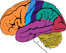
Each cerebral hemisphere can be divided into sections, or lobes, each of which specializes in different functions. To understand each lobe and its specialty, we will take a tour of the cerebral hemispheres.
Frontal lobes

The two frontal lobes lie directly behind the forehead. When you plan a schedule, imagine the future, or use reasoned arguments, these two lobes do much of the work. One of the ways the frontal lobes seem to do these things is by acting as short-term storage sites, allowing one idea to be kept in mind while other ideas are considered.
Motor cortex
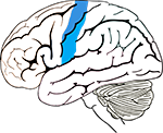
In the back portion of each frontal lobe is a motor cortex, which helps plan, control, and execute voluntary movement, like moving your arm or kicking a ball.
Parietal lobes

When you enjoy a good meal—the taste, smell, and texture of the food—two sections behind the frontal lobes called the parietal lobes are at work. The parietal lobes also support reading and arithmetic.
Somatosensory cortex

The forward parts of these lobes, just behind the motor areas, are the somatosensory cortex. These areas receive information about temperature, taste, touch, and movement from the rest of the body.
Occipital lobes

As you look at the words and pictures on this page, two areas at the back of the brain are at work. These lobes, called the occipital lobes, process images from the eyes and link that information with images stored in memory. Damage to the occipital lobes can cause blindness.
Temporal lobes

The last lobes on our tour of the cerebral hemispheres are the temporal lobes, which lie in front of the visual areas and nest under the parietal and frontal lobes. Whether you appreciate symphonies or rock music, your brain responds through the activity of these lobes. At the top of each temporal lobe is an area responsible for receiving information from the ears. The underside of each temporal lobe plays a crucial role in forming and retrieving memories, including those associated with music. Other parts of this lobe integrate memories and sensations of taste, sound, sight, and touch.
The Inner Brain
Deep within the brain, hidden from view, lie structures that are the gatekeepers between the spinal cord and the cerebral hemispheres. These structures not only determine our emotional state, but they also modify our perceptions and responses and allow us to initiate movements without thinking about them. Like the lobes in the cerebral hemispheres, the structures described below come in pairs: Each is duplicated in the opposite half of the brain.

The hypothalamus, about the size of a pearl, directs a multitude of important functions. It wakes you up in the morning and gets the adrenaline flowing during a test or job interview. The hypothalamus is also an important emotional center, controlling the chemicals that make you feel exhilarated, angry, or unhappy. Near the hypothalamus lies the thalamus, a major clearinghouse for information going to and from the spinal cord and the cerebrum.
An arching tract of nerve cells leads from the hypothalamus and the thalamus to the hippocampus. This tiny nub acts as a memory indexer—sending memories out to the appropriate part of the cerebral hemisphere for long-term storage and retrieving them when necessary. The basal ganglia (not shown) are clusters of nerve cells surrounding the thalamus. They are responsible for initiating and integrating movements. Parkinson’s disease, which results in tremors, rigidity, and a stiff, shuffling walk, affects the nerve cells in the basal ganglia.
The Neuron
The brain and the rest of the nervous system are composed of many different types of cells, but the primary functional unit is a cell called the neuron. All sensations, movements, thoughts, memories, and feelings are the result of signals that pass through neurons. Neurons consist of three parts: the cell body, dendrites, and the axon.
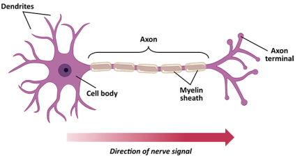
The cell body contains the nucleus, where most of the molecules that the neuron needs to survive and function are manufactured. Dendrites extend out from the cell body like the branches of a tree and receive messages from other nerve cells. Signals then pass from the dendrites through the cell body and travel away from the cell body down an axon to another neuron, a muscle cell, or cells in some other organ.
The neuron is usually surrounded by many support cells. Some types of cells wrap around the axon to form an insulating myelin sheath. Myelin is a fatty molecule which provides insulation for the axon and helps nerve signals travel faster and farther. Axons may be very short, such as those that carry signals from one cell in the cortex to another cell less than a hair’s width away. Other axons may be very long, such as those that carry messages from the brain all the way down the spinal cord.
The Synapse

Scientists have learned a great deal about neurons by studying the synapse – the place where a signal passes from one neuron to another. When the signal reaches the end of the axon, it stimulates the release of tiny sacs called vesicles. These vesicles release chemicals known as neurotransmitters into the synaptic cleft. The neurotransmitters cross the synapse and attach to receptors on the neighboring cell. These receptors can change the properties of the receiving cell. If the receiving cell is also a neuron, the signal can continue the transmission to the next cell.
Links to Learning
- Hand-Brain Model (Youtube)- Learn to use your fist to model the brain and use it to discuss the difference between the logical brain vs the emotional brain.
- Debunking Myths about the Human Brain (from the Global Council on Brain Health)
- Mapping the Brain (Nova scienceNow)- In this interactive activity from the NOVA scienceNOW website, learn about several brain mapping techniques: MRI, fMRI, PET, MEG, DTI, and probabilistic.
- Understanding Your Brain to Help You Learn Better (by Frontiers for Young Minds)
Review & Practice
Distinguish between the cerebral cortex and the cerebrum.
Critical Thinking
- Which part(s) of your brain are overactive? Underactive? Explain.
- Hypothetically speaking, choose a region of the brain, and create a marketing advertisement to sell its attributes.
- What structure of the brain is most responsible for stress and anxiety? Why do you think this structure of the brain is so reactive?31
This chapter was adapted from The National Institute of Neurological Disorders and Stroke (NINDS). (2024). Brain Basics: Know Your Brain | National Institute of Neurological Disorders and Stroke.
References
Biswas-Diener, R. (2025). The brain and nervous system. In R. Biswas-Diener & E. Diener (Eds), Noba textbook series: Psychology. Champaign, IL: DEF publishers. Retrieved from http://noba.to/4hzf8xv6 on January 5, 2025.
Hovden, J., Nguyen, A., & Ortega, A. (2020). Learning to Learn. Pp. 165-176. Retrieved from Couns142_version1.pdf on January 5, 2025.
National Institute of Neurological Disorders and Stroke (NINDS). (2024). Brain Basics: Know Your Brain. Retrieved from https://www.ninds.nih.gov/health-information/public-education/brain-basics/brain-basics-know-your-brain on January 5, 2025
Media Attributions
- Figure 1.11 © National Institute of Neurological Disorders and Stroke is licensed under a Public Domain license
- Figure 1.5 Frontal lobe © National Institute of Neurological Disorders and Stroke is licensed under a Public Domain license
- Figure 1.6 Motor cortex © National Institute of Neurological Disorders and Stroke is licensed under a Public Domain license
- Figure 1.7 Parietal lobes © National Institute of Neurological Disorders and Stroke is licensed under a Public Domain license
- Figure 1.8 Somatosensory cortex © National Institute of Neurological Disorders and Stroke is licensed under a Public Domain license
